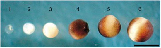
Dumont 标准依据直径、色素沉着与形态特征,将爪蟾卵母细胞分为六个时期(I–VI)。正确分期是微注射、蛋白表达与电生理实验稳定可重复的前提。The Dumont system classifies Xenopus oocytes into six stages (I–VI) by diameter, pigmentation and morphology — the basis for reproducible injection, expression and electrophysiology.
分期标准Staging
| 分期Stage | 直径Diameter | 主要特征Features |
|---|---|---|
| I | 50–100 μm | 胞体透明,无色素Transparent, no pigment |
| II | 300–450 μm | 透明或淡白色Transparent / pale |
| III | 450–600 μm | 表面出现均匀色素Uniform pigment appears |
| IV | 600–1000 μm | 出现深色动物半球与白色植物半球Animal/vegetal hemispheres form |
| V | 1000–1200 μm | 两半球界限清晰,动物半球深黑发亮Clear hemisphere border |
| VI | 1200–1300 μm | 无色素带分隔,体积最大Unpigmented band, largest |
实验选用建议Selection
- 微注射(mRNA/cRNA):推荐 VI 期——体积最大(注射量可至 100 nL)、结构完整、翻译效率高。Injection (mRNA/cRNA): prefer stage VI — largest volume (up to 100 nL), intact, high translation.
- 电生理(电压钳):推荐 II–III 期——体积小、电容低、噪声低、稳定时间短。Electrophysiology: prefer stage II–III — lower capacitance and noise.
应结合直径测量与形态观察进行分期;保存于含抗生素的 0.1× MMR 中;大细胞注射需控制针径与压力。Combine diameter and morphology; keep in 0.1× MMR with antibiotics; control needle size and pressure.
参考文献References
- Dumont JN (1972) Oogenesis in Xenopus laevis (Daudin). I. Stages of oocyte development in laboratory maintained animals. J Morphol 136:153–180.
- Goldin AL (1992) Maintenance of Xenopus laevis and oocyte injection. Methods Enzymol 207:266–279.
- Sigel E (1990) Use of Xenopus oocytes for the functional expression of plasma membrane proteins. J Membr Biol 117(3):201–221.