爪蟾卵母细胞是最常用的功能表达系统之一。将体外转录的 cRNA 微注射到卵母细胞的细胞质中,目标蛋白通常次日即可在细胞膜上检测到功能性表达,可直接用于双电极电压钳(TEVC)电生理记录。Xenopus oocytes are one of the most widely used heterologous expression systems. Microinjecting in vitro-transcribed cRNA into the cytoplasm yields functional membrane-protein expression detectable by the next day, ready for two-electrode voltage clamp (TEVC) electrophysiology.
1. cRNA 准备1. cRNA preparation
- 以线性化质粒为模板,用 mMessage mMachine 等试剂盒体外转录合成 cRNA;加约 300 nt poly(A) 尾以提高翻译效率与稳定性。In vitro transcription from linearised plasmid using kits such as mMessage mMachine (Ambion); add ~300 nt poly(A) tail to boost translation efficiency and stability.
- 产物在含 Radiant Red 等 RNA 染料的琼脂糖凝胶上定量;多亚基蛋白按摩尔比混合后溶于无 RNase 水,−80 ℃ 保存。Quantify on agarose gel with RNA stain; mix multi-subunit cRNAs at molar ratio in RNase-free water; store at −80 ℃.
2. 卵巢取出与卵母细胞分离2. Ovary removal & oocyte isolation
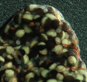
- MS-222 麻醉后手术取出卵巢袋(包裹在含血管的结缔组织中),置于含 1× MBS-H(无 Ca²⁺)的培养皿。After MS-222 anaesthesia, surgically excise the ovary sacs (embedded in vascularised connective tissue); transfer to 1× MBS-H (Ca²⁺-free).
- 用铂环(或镊子)机械分离单个卵母细胞(直径 1.0–1.2 mm);或用胶原酶(2 mg/mL,1–2 h,20 ℃)酶解分离,彻底洗去胶原酶后使用。Separate single oocytes (1.0–1.2 mm) mechanically with a platinum loop or forceps; alternatively, enzymatic dissociation with collagenase (2 mg/mL, 1–2 h, 20 ℃) followed by thorough washing.
- 选取 V–VI 期健康卵母细胞,排列于内衬尼龙网的塑料网格上,注射前在 0.1× MMR(含 50 μg/mL 庆大霉素)中 18 ℃ 预孵育 ≥2 h。Select healthy stage V–VI oocytes; arrange on a nylon mesh grid; pre-incubate in 0.1× MMR with 50 μg/mL gentamicin at 18 ℃ for ≥2 h before injection.
3. 去卵泡处理3. Defolliculation
- 胶原酶分离的卵母细胞仍可能残留卵泡细胞层,影响电压钳记录。可用高渗 EGTA 溶液(82.5 mM KCl, 1 mM MgCl₂, 10 mM HEPES, 10 mM EGTA, pH 7.4)轻柔处理去除。Collagenase-dissociated oocytes may retain follicle-cell layers that interfere with TEVC. Remove residual follicle cells with hypertonic EGTA solution (82.5 mM KCl, 1 mM MgCl₂, 10 mM HEPES, 10 mM EGTA, pH 7.4).
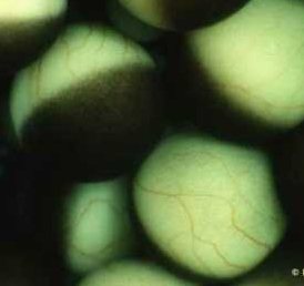
- 去卵泡后再次在 0.1× MMR 中充分洗涤,选取表面光滑、两半球界限清晰的健康 VI 期细胞用于注射。Wash thoroughly in 0.1× MMR after defolliculation; select healthy stage VI oocytes with smooth surface and clear hemisphere border for injection.
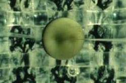
4. 注射参数4. Injection parameters
| 参数Parameter | 推荐值Value | 说明Note |
|---|---|---|
| 注射量Volume | ~50 nL | 可至 100 nL,不超过卵母细胞体积的 10%Up to 100 nL; do not exceed 10% of oocyte volume |
| 针尖直径Tip diameter | ~15 μm | 太细易堵塞,太粗损伤细胞Too narrow clogs; too wide damages oocyte |
| 注射时间Duration | ~5 s | 缓慢注射减少损伤Slow injection minimises damage |
| 注射部位Site | 赤道偏动物极,细胞质Equatorial-animal cytoplasm | 避开核区,避免核注射Avoid nuclear region |
| 孵育温度Incubation | 18–20 ℃ | 注射后 0.1× MMR 中 16–48 hIn 0.1× MMR for 16–48 h post-injection |
5. 功能检测5. Functional readout
- 表达蛋白的功能通常在注射后次日(16–24 h)可测量;最大表达高峰在 2–4 天。Functional expression is typically measurable 16–24 h post-injection; peak expression occurs at 2–4 days.
- 双电极电压钳(TEVC):两根尖端 ~1 μm 的微电极分别插入卵母细胞,一根固定膜电位,另一根记录穿膜离子电流。TEVC: two ~1 μm microelectrodes are inserted into the oocyte — one clamps membrane voltage, the other records transmembrane ionic current.
- 转运蛋白功能可用放射性同位素(¹⁴C、³H 标记底物)或荧光底物进行摄取实验定量。Transporter function can be quantified by uptake assays with radiolabelled (¹⁴C, ³H) or fluorescent substrates.
必须设对照:非注射卵母细胞(uninjected)与注射水的卵母细胞(water-injected)均需同步记录,以扣除内源性电流背景。多亚基蛋白表达时需按照合适摩尔比混合各亚基 cRNA。Always include controls: uninjected and water-injected oocytes must be recorded in parallel to subtract endogenous current background. For multi-subunit proteins, mix subunit cRNAs at the appropriate molar ratio.
参考文献References
- Sigel E (1987) Properties of single sodium channels translated by Xenopus oocytes after injection with messenger ribonucleic acid. J Physiol (Lond) 386:73–90.
- Sigel E, et al. (1990) The effect of subunit composition of rat brain GABAA receptors on channel function. Neuron 5(6):703–711.
- Sigel E (1990) Use of Xenopus oocytes for the functional expression of plasma membrane proteins. J Membr Biol 117(3):201–221.